Boyle 2017 MiP2017
| Type 2 diabetes causes systemic skeletal muscle complex I mitochondrial dysfunction in chronic heart failure patients. |
Link: MiP2017
Garnham JO, Bowen TS, Witte KK, Boyle JP (2017)
Event: MiP2017
Chronic heart failure (CHF) is characterised by exercise intolerance, which is exacerbated in patients with concomitant type 2 diabetes (CHF+DM) independent of left ventricular ejection fraction. This may be attributed to non-cardiac alterations, with greater skeletal muscle mitochondrial dysfunction a potential underlying mechanism. However, previous studies in CHF have used single isolated biopsies from peripheral locomotor muscles, which are susceptible to detraining, thereby confounding the results.
We assessed the hypothesis that CHF+DM patients are characterised by systemic skeletal muscle mitochondrial dysfunction by taking biopsies from two muscles.
Skeletal muscle biopsies were sampled consecutively from vastus lateralis and pectoralis major from age-matched healthy controls (n=6), CHF (n=12), and CHF+DM patients (n=7). Mitochondrial oxygen flux of permeabilized fibres was subsequently assessed using a standard substrate, uncoupler and inhibitor titration protocol to selectively measure isolated components of the electron transfer chain.
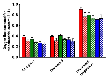
After correcting for mitochondrial content, we found a significant main effect for group for complex I oxygen flux, F (2, 22) = 4.16, p = 0.029, with a significant difference between CHF+DM and healthy controls in both muscles (Figure 1). In contrast, CHF patients alone were not different to controls.
In conclusion, CHF+DM patients present with a systemic skeletal muscle mitochondrial dysfunction, which is not apparent in CHF patients. These findings provide an initial rationale for why exercise intolerance is exacerbated in CHF+DM.
• Bioblast editor: Kandolf G
• O2k-Network Lab: UK Leeds Peers C
Labels: MiParea: Respiration Pathology: Cardiovascular, Diabetes
Organism: Human Tissue;cell: Skeletal muscle Preparation: Permeabilized tissue
Coupling state: ET-pathway"ET-pathway" is not in the list (LEAK, ROUTINE, OXPHOS, ET) of allowed values for the "Coupling states" property.
Pathway: N, S
HRR: Oxygraph-2k
Figure 1
Figure 1: Skeletal muscle oxygen flux corrected for mitochondrial content (A.U.) measured in 3 different respiratory states from age-matched healthy controls (red), CHF patients (green), and CHF+DM patients (blue). Filled bars indicate pectoralis major samples. Striped bars indicate vastus lateralis samples. Data are expressed as mean ± SEM. Between group differences were assessed using a 2-way mixed analysis of variance (ANOVA) and Bonferroni post hoc analyses.
Affiliations
Division Cardiovascular Diabetes Research, Leeds Inst Cardiovascular Metabolic Medicine, Fac Medicine Health, Univ Leeds, UK. - [email protected]


