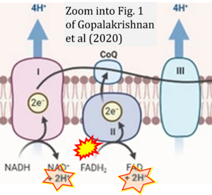
Gopalakrishnan 2020 Sci Rep
| Gopalakrishnan S, Mehrvar S, Maleki S, Schmitt H, Summerfelt P, Dubis AM, Abroe B, Connor TB Jr, Carroll J, Huddleston W, Ranji M, Eells JT (2020) Photobiomodulation preserves mitochondrial redox state and is retinoprotective in a rodent model of retinitis pigmentosa. Sci Rep 10:20382. https://doi.org/10.1038/s41598-020-77290-w |
Gopalakrishnan S, Mehrvar S, Maleki S, Schmitt H, Summerfelt P, Dubis AM, Abroe B, Connor TB Jr, Carroll J, Huddleston W, Ranji M, Eells JT (2020) Sci Rep
Abstract: Photobiomodulation (PBM) by far-red (FR) to near-infrared (NIR) light has been demonstrated to restore the function of damaged mitochondria, increase the production of cytoprotective factors and prevent cell death. Our laboratory has shown that FR PBM improves functional and structural outcomes in animal models of retinal injury and retinal degenerative disease. The current study tested the hypothesis that a brief course of NIR (830 nm) PBM would preserve mitochondrial metabolic state and attenuate photoreceptor loss in a model of retinitis pigmentosa, the P23H transgenic rat. P23H rat pups were treated with 830 nm light (180 s; 25 mW/cm2; 4.5 J/cm2) using a light-emitting diode array (Quantum Devices, Barneveld, WI) from postnatal day (p) 10 to p25. Sham-treated rats were restrained, but not treated with 830 nm light. Retinal metabolic state, function and morphology were assessed at p30 by measurement of mitochondrial redox (NADH/FAD) state by 3D optical cryo-imaging, electroretinography (ERG), spectral-domain optical coherence tomography (SD-OCT), and histomorphometry. PBM preserved retinal metabolic state, retinal function, and retinal morphology in PBM-treated animals compared to the sham-treated group. PBM protected against the disruption of the oxidation state of the mitochondrial respiratory chain observed in sham-treated animals. Scotopic ERG responses over a range of flash intensities were significantly greater in PBM-treated rats compared to sham controls. SD-OCT studies and histological assessment showed that PBM preserved the structural integrity of the retina. These findings demonstrate for the first time a direct effect of NIR PBM on retinal mitochondrial redox status in a well-established model of retinal disease. They show that chronic proteotoxic stress disrupts retinal bioenergetics resulting in mitochondrial dysfunction, and retinal degeneration and that therapies normalizing mitochondrial metabolism have considerable potential for the treatment of retinal degenerative disease.
• Bioblast editor: Gnaiger E
Correction: FADH2 and Complex II
- FADH2 is shown as the substrate feeding electrons into Complex II (CII). This is wrong and requires correction - for details see Gnaiger (2024).
- Gnaiger E (2024) Complex II ambiguities ― FADH2 in the electron transfer system. J Biol Chem 300:105470. https://doi.org/10.1016/j.jbc.2023.105470 - »Bioblast link«
Hydrogen ion ambiguities in the electron transfer system
Communicated by Gnaiger E (2023-10-08) last update 2023-11-10
- Electron (e-) transfer linked to hydrogen ion (hydron; H+) transfer is a fundamental concept in the field of bioenergetics, critical for understanding redox-coupled energy transformations.
- However, the current literature contains inconsistencies regarding H+ formation on the negative side of bioenergetic membranes, such as the matrix side of the mitochondrial inner membrane, when NADH is oxidized during oxidative phosphorylation (OXPHOS). Ambiguities arise when examining the oxidation of NADH by respiratory Complex I or succinate by Complex II.
- Oxidation of NADH or succinate involves a two-electron transfer of 2{H++e-} to FMN or FAD, respectively. Figures indicating a single electron e- transferred from NADH or succinate lack accuracy.
- The oxidized NAD+ is distinguished from NAD indicating nicotinamide adenine dinucleotide independent of oxidation state.
- NADH + H+ → NAD+ +2{H++e-} is the oxidation half-reaction in this H+-linked electron transfer represented as 2{H++e-} (Gnaiger 2023). Putative H+ formation shown as NADH → NAD+ + H+ conflicts with chemiosmotic coupling stoichiometries between H+ translocation across the coupling membrane and electron transfer to oxygen. Ensuring clarity in this complex field is imperative to tackle the apparent ambiguity crisis and prevent confusion, particularly in light of the increasing number of interdisciplinary publications on bioenergetics concerning diagnostic and clinical applications of OXPHOS analysis.


